
Background
Subject motion during during external beam radiotherapy has been problematic since its introduction as a clinical therapeutic modality. This is primarily due to the prolonged time required for radiation delivery, which is far longer than the timescale of most types of physiological motion, including gastrointestinal peristalsis, cardiac and respiratory motion, and involuntary bulk motion. The figure below shows 4D-MRI videos of some of these types of motion for different anatomical locations. Here you can clearly observe large tissue displacements as a result of the physiological motion. These diplacements ultimately limit the accuracy of the radiation delivery in two distinct ways:
- Motion during the pre-treatment MR imaging results into motion artefacts in the MR images. These motion artefacts reduce the delineation accuracy and therefore requires motion corrected image reconstruction.
- Motion during the radiation delivery deposites the radiation dose in the wrong location. This requires fast 3D motion estimation to adapt the radiation beam in real-time.
Therefore, the research within this project focuses on real-time 3D motion estimation and how to use this motion information for motion corrected image reconstruction.
RESOLVE
RESOLVE stands for “Enabling MR guided radiothErapy of moving abdominal tumorS and Organs by development of a dense eLement receiVer array and highly undersampled, parallEl MRI”. In this project we are developing new and faster motion resolved MRI methods that will provide the means to monitor 3D dose deposition in moving tissue or even lock the radiation beam to the moving tumor. This unlocks the potential of MRL for radiation treatment of abdominal tumors resulting in more effective radiation treatment of abdominal cancers and lower toxicity of healthy organs-at-risk.
This work is part of the research program HTSM with project number 15354, which is (partly) financed by the Netherlands Organization for Scientific Research (NWO) and PHILIPS healthcare.
MR-MOTUS
MR-MOTUS is a framework to estimate 3D non-rigid motion-fields directly from k-space data and a reference image, and the acronym stands for Model-based Reconstruction of MOTion from Undersampled Signal. Recently, the MR-Linac was introduced as a hybrid between an MR-scanner and a radiotherapy linear accelator (Linac), which has the ultimate potential to perform real-time adaptive intra-fraction MR-guided abdominal radiotherapy. A requirement for this a sufficiently high framerate of 5 Hz for both reconstruction and acquisition of data, which is typically very challenging due to the relatively slow imaging speed of MRI and long reconstruction times for advanced techniques. The MR-MOTUS framework was designed to fill in this gap, and estimate non-rigid 3D motion at more than 5 Hz, including both reconstruction and data acquisition.
The MR-MOTUS forward model relates motion-fields and a reference image to non-Cartesian k-space data. The general idea is that a reference image is reconstructed before the treatment starts, and motion-fields are subsequently reconstructed from highly undersampled k-space data with a model-based reconstruction. The main advantage of the framework is that it enables to extract motion directly from k-space data, rather than indirectly through image reconstruction. The framework is therefore very flexible in terms of temporal resolution and acquisition trajectories, and is not directly limited by the achievable temporal resolution of MR-images.
Figure 2: Low-rank MR-MOTUS applied to rapid 2D cine MRI with golden angle radial sampling.
Figure 3: 3D time-resolved cine reconstruction capturing breathing motion at a temporal resolution of ~100 ms using CMR-MOTUS..
This work is funded by the NWO VENI award of Dr. Alessandro Sbrizzi, grant number 15115.
Related publications:
- Huttinga, N. R., Bruijnen, T., van den Berg, C. A., & Sbrizzi, A. (2023). Gaussian Processes for real-time 3D motion and uncertainty estimation during MR-guided radiotherapy. Medical Image Analysis, 88, 102843.
- Huttinga, N. R., Bruijnen, T., Van Den Berg, C. A., & Sbrizzi, A. (2021). Real-time non-rigid 3D respiratory motion estimation for MR-guided radiotherapy using MR-MOTUS. IEEE Transactions on Medical Imaging, 41(2), 332-346.
- Huttinga NRF, Bruijnen T, van den Berg CAT, Sbrizzi A. Nonrigid 3D motion estimation at high temporal resolution from prospectively undersampled k-space data using low-rank MR-MOTUS. Magn Reson Med. 2020 Nov 10. doi: 10.1002/mrm.28562. Epub ahead of print. PMID: 33169888. (paper). (code)
- Huttinga NRF, van den Berg CAT, Luijten PR, Sbrizzi A. MR-MOTUS: model-based non-rigid motion estimation for MR-guided radiotherapy using a reference image and minimal k-space data. Phys Med Biol 2020 Jan 10. doi: 10.1088/1361-6560/ab554a. PMID: 31698354. (paper)
Motion-Free MRI
Several patient groups (e.g. brain tumor, epilepsy, multiple sclerosis, stroke, neurogenerative disorders and children in general) rely heavily on MRI scans for diagnosis and periodic check-ups. At the same time, repeat scans due to motion artefacts are more common amongst these groups and research shows that more than 15% of MRI scans needs to be repeated. Children usually undergo complex anaesthesia procedures to prevent them from moving. Our goal is to dramatically reduce the need for scans repetitions and/or anaesthesia by changing the way images are constructed from conventional MRI protocols. We aim to achieve this by developing an algorithm for retrospective motion correction that does not require modifications to current clinical MRI acquisition protocols, leading to potentially fast and broad clinical deployment.
Our Motion-Free MRI algorithm is based on a rigid motion model and assumes that at least one of the contrasts is motion-free. The method simultaneously reconstructs the images from MRI data and estimates the parameters of the rigid motion model. It corrects the corrupted sequences by minimizing data-fit and maximising image similarity with respect to the reference contrast by means of structural-guided-Total-Variation (sgTV). The algorithm consists of three main building blocks: (1) a measurement model to predict the measured MR data for given contrast images, (2) a 3D motion model which rigidly deforms the images according to its parameters, and (3) a measure of image similarity (sgTV) which relates the anatomical structures of the images being reconstructed.
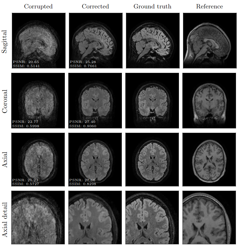
Figure 3: example of the 3D results that can be achieved with our Motion-Free-MRI technology (version 1). Slices from a 3D motion-corrupted T2-flair image are shown (first column) along with a 3D motion-free T1 image (last column) which is used as reference. The corrected T2-flair image (second column) clearly shows the expected anatomical features. The indicated PSNR (in dB) and SSIM (on a scale 0 – 1 with 1 being the best achievable similarity) are quantitative indicators of image quality with respect to the known (motion-free) ground-truth T2-flair (third column).
Related publications:
- Rizzuti, G., Schakel, T., Huttinga, N. R., Dankbaar, J. W., van Leeuwen, T., & Sbrizzi, A. (2023). Towards retrospective motion correction and reconstruction for clinical 3D brain MRI protocols with a reference contrast. Magnetic Resonance Materials in Physics, Biology and Medicine (MAGMA) [In press] arXiv preprint arXiv:2301.01106.
- Rizzuti, G., Sbrizzi, A., & Van Leeuwen, T. (2022). Joint retrospective motion correction and reconstruction for brain MRI with a reference contrast. IEEE Transactions on Computational Imaging, 8, 490-504.
Collaborating partners:

People


Max van Riel

Maarten Terpstra

Thomas Olausson

Matteo Maspero

Alessandro Sbrizzi
